.png)

阴道毛滴虫(Trichomonas vaginalis)
物种名:阴道毛滴虫
拉丁学名:Trichomonas vaginalis
分类学地位: 真核生物界Eukaryota;副基体门Parabasalia;
毛滴虫纲Trichomonadea毛滴虫目Trichomonadida;
毛滴虫科Trichomonadidae;毛滴虫属Trichomonas
阴道毛滴虫(Trichomonas vaginalis)是一种寄生于人类泌尿生殖道的鞭毛虫,主要引起滴虫性阴道炎(trichomoniasis),是全球最常见的非病毒性性传播感染之一。该虫可通过性接触传播,也可通过间接接触污染物品传播,女性感染率高于男性,且常与不良妊娠结局和HIV传播风险增加相关。
1.1生物学特性
1.1.1培养特征
阴道毛滴虫为厌氧或微需氧生物,最适生长温度为35-37℃,最适pH为5.5-6.0。可在Diamond’s TYM培养基、InPouch TV培养系统等中生长,培养48-72小时后可见虫体增殖,呈梨形或椭圆形,具有鞭毛和波动膜[1]。
1.1.2形态学特征
阴道毛滴虫呈梨形,大小约为10-20 μm,具4根前鞭毛和1根后鞭毛,后鞭毛与波动膜相连,无包囊阶段,仅有滋养体形态。虫体可通过鞭毛和波动膜进行快速运动,吉姆萨染色或巴氏涂片可见典型结构(图1)[2]。
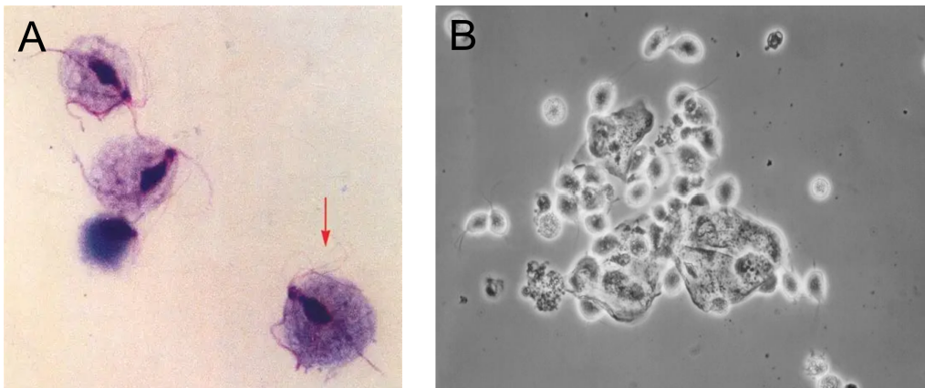
图1 阴道毛滴虫照片
A:阴道毛滴虫显微照片(×1000)[3],B:阴道毛滴虫显微镜图[4]
1.1.3生化特征
阴道毛滴虫缺乏线粒体,依赖氢化酶体进行能量代谢,能发酵多种糖类产生乙酸、乳酸等有机酸。其表面富含半胱氨酸蛋白酶、黏附蛋白等毒力因子,能降解宿主细胞外基质,促进虫体定植[5]。
1.1.4分子生物学特征
阴道毛滴虫基因组大小约160 Mb,编码约60000个基因,具有高度重复序列和大量横向转移基因。其毒力相关基因包括编码黏附素(AP65、AP33等)的基因、编码半胱氨酸蛋白酶(CPs)的基因以及调控运动的鞭毛蛋白基因等[6]。
1.2分布、传播与致病性
1.2.1分布与传播
阴道毛滴虫全球分布,尤其在卫生条件较差和性健康意识薄弱地区感染率较高。主要通过性接触传播,也可通过共用毛巾、浴具等间接途径传播。女性感染者多见,男性常为无症状携带者[7]。
1.2.2致病性
感染后可引起滴虫性阴道炎,典型症状包括阴道分泌物增多(黄绿色、泡沫状)、外阴瘙痒、排尿困难等。男性感染可导致非淋菌性尿道炎。该虫可破坏生殖道上皮屏障,增加HIV等其他性传播疾病的感染风险[8]。
阴道毛滴虫的致病机制主要与虫体黏附于宿主细胞表面有关,其黏附涉及四种黏附蛋白(AP65、AP51、AP33、AP23)及半胱氨酸蛋白酶;同时,虫体可通过细胞黏附、溶血、细胞脱落因子及逃避宿主免疫系统等多种机制致病。其中,虫体分泌的蛋白酶可降解宿主免疫球蛋白,从而帮助寄生虫逃避宿主免疫系统的识别与清除[9]。
1.3检测方法
(1)显微镜检查:取阴道分泌物或尿道分泌物直接涂片,显微镜下观察活动虫体,方法简便但灵敏度较低[10]。
(2)分子生物学方法:以阴道毛滴虫基因组中特异性保守序列A6p为检测靶点,建立基于PCR的分子生物学检测方法。首先从临床样本(如阴道拭子)中提取总DNA作为模板,加入特异性引物对TVA5和TVA6进行PCR扩增,其中TVA5序列为5′-GATCATGTTCTATCTTTTCA-3′,TVA6序列为5′-GATCACCACCTTAGTTTACA-3′。反应条件为94℃预变性7 min,然后进行30-45个循环(94℃变性1 min、47℃退火1 min、67℃延伸1 min),最后67℃延伸7 min。扩增产物经琼脂糖凝胶电泳和溴化乙锭染色后,若在紫外灯下观察到102 bp的特异性条带,即可判定为阳性。该引物对与人体DNA及其他多种原虫、细菌、真菌无交叉反应,具有高度敏感性和特异性[11]。
(4)抗原检测:采用免疫层析或ELISA法检测虫体特异性抗原,操作简便,适用于快速诊断[12]。
1.4典型案例
美国一项基于全国代表性样本(NHANES 2001-2004)的研究显示,阴道毛滴虫感染率在非西班牙裔黑人女性中显著高于其他种族人群——非西班牙裔黑人女性患病率为13.3%,是非西班牙裔白人(1.3%)和墨西哥裔美国女性(1.8%)的10.3倍;同时,该感染与早产、低出生体重婴儿分娩等不良妊娠/不良健康结局相关[13]。一项在非洲(肯尼亚蒙巴萨)女性中开展的前瞻性研究显示,感染阴道毛滴虫与HIV-1感染获得风险增加1.52倍(95% CI 1.04–2.24)相关,研究认为治疗和预防阴道毛滴虫感染可能降低HIV-1感染风险[14]。
1.5防治对策
预防滴虫感染需加强性健康教育,推广使用安全套,避免不洁性行为。感染者及其性伴侣应同时接受治疗,常用药物为甲硝唑或替硝唑。注意个人卫生,避免共用洗浴用品,定期进行妇科检查,尤其是有症状或高风险人群[15]。
参考文献
[1] Menezes CB, Frasson AP, Tasca T. Optimal conditions for the continuous axenic cultivation of Trichomonas vaginalis. Parasitology, 2016, 143: 1664-1671.
[2] Ryan KJ. Sherris Medical Microbiology. McGraw Hill, 2010.
[3] Garcia K, Corona S, Rodriguez A et al. Comparative morphological analysis of Trichomonas vaginalis adherence to host cells using light and scanning electron microscopy. Parasitology Research, 2021, 120: 987-995.
[4] Centers for Disease Control and Prevention. Trichomonas vaginalis in wet mount vaginal discharge specimen. Public Health Image Library, Image ID: 14500.
[5] Hirt RP. Trichomonas vaginalis pathogenicity: update and perspectives. Archives of Medical Research, 2015, 46: 33-39.
[6] Carlton JM. Draft genome sequence of the sexually transmitted pathogen Trichomonas vaginalis. Science, 2007, 315: 207-212.
[7] Rowley J, Vander Hoorn S, Korenromp E et al. Chlamydia, gonorrhoea, trichomoniasis and syphilis: global prevalence and incidence estimates, 2016. Bulletin of the World Health Organization, 2019, 97: 548-562.
[8] Kissinger P. Trichomonas vaginalis: a review of epidemiologic, clinical and treatment issues. BMC Infectious Diseases, 2015, 15: 307.
[9] Adegbaju, A., Morenikeji et al. Cytoadherence and pathogenesis of Trichomonas vaginalis. Scientific Research and Essays, 2008, Vol.3: 132-138.
[10] Van Der Pol B. Clinical and laboratory testing for Trichomonas vaginalis infection. Journal of Clinical Microbiology, 2016, 54: 7-12.
[11] Riley DE, Roberts MC, Takayama T et al. Development of a polymerase chain reaction-based diagnosis of Trichomonas vaginalis. Journal of Clinical Microbiology, 1992, 30: 465-472.
[12] Huppert JS, Mortensen JE, Reed JL et al. Rapid antigen testing compares favorably with transcription-mediated amplification assay for the detection of Trichomonas vaginalis in young women. Clinical Infectious Diseases, 2007, 45: 194-198.
[13] Sutton M., Sternberg M., Koumans E.H. et al. The prevalence of Trichomonas vaginalis infection among reproductive-age women in the United States, 2001-2004. Clinical Infectious Diseases, 2007, Vol.45: 1319-1326.
[14] McClelland, Scott R. 1,2 et al. Infection with Trichomonas vaginalis Increases the Risk of HIV-1 Acquisition. Journal of Infectious Diseases, 2007, Vol.195: 698-702.
[15] Workowski KA, Bachmann LH, Chan PA et al. Sexually Transmitted Infections Treatment Guidelines, 2021. MMWR Recommendations and Reports, 2021, 70: 1-187.